Reseptor warna: Perbedaan antara revisi
k Perubahan kosmetik tanda baca |
Add 2 books for Wikipedia:Pemastian (20210209)) #IABot (v2.0.8) (GreenC bot |
||
| Baris 28: | Baris 28: | ||
|year = 2000 |
|year = 2000 |
||
|title = Principles of Neural Science |
|title = Principles of Neural Science |
||
|url = https://archive.org/details/principlesneural00kand_658 |
|||
|edition = 4th |
|edition = 4th |
||
|pages = [https://archive.org/details/principlesneural00kand_658/page/n436 507]–513 |
|||
|pages = 507–513 |
|||
|publisher = McGraw-Hill |
|publisher = McGraw-Hill |
||
|location = New York |
|location = New York |
||
| Baris 77: | Baris 78: | ||
| year = 2004 |
| year = 2004 |
||
| title = The Reproduction of Colour |
| title = The Reproduction of Colour |
||
| url = https://archive.org/details/reproductionofco0000hunt |
|||
| edition = 6th |
| edition = 6th |
||
| pages = [https://archive.org/details/reproductionofco0000hunt/page/11 11]–12 |
|||
| pages = 11–12 |
|||
| publisher = Wiley–IS&T Series in Imaging Science and Technology |
| publisher = Wiley–IS&T Series in Imaging Science and Technology |
||
| location = Chichester UK |
| location = Chichester UK |
||
Revisi per 11 Februari 2021 10.13
Artikel ini perlu diterjemahkan dari bahasa Inggris ke bahasa Indonesia. Artikel ini ditulis atau diterjemahkan secara buruk dari Wikipedia bahasa Inggris. Jika halaman ini ditujukan untuk komunitas bahasa Inggris, halaman itu harus dikontribusikan ke Wikipedia bahasa Inggris. Lihat daftar bahasa Wikipedia. Artikel yang tidak diterjemahkan dapat dihapus secara cepat sesuai kriteria A2. Jika Anda ingin memeriksa artikel ini, Anda boleh menggunakan mesin penerjemah. Namun ingat, mohon tidak menyalin hasil terjemahan tersebut ke artikel, karena umumnya merupakan terjemahan berkualitas rendah. |
| Neuron: Cone cell | |
|---|---|
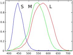
 Spektrum responsiviti pada sel kerucut manusia tipe S, M, dan L | |
| Fungsi | Melihat warna |
| ID NeuroLex | sao1103104164 |

Reseptor warna atau sering juga disebut sel kerucut (Inggris: cone cell) adalah sel penerima sinar di dalam retina mata yang bertanggung jawab terhadap penglihatan warna. Sel kerucut akan bekerja dengan baik pada kondisi yang cukup terang. Sebagai lawannya, sel batang akan bekerja dengan baik pada cahaya yang redup.
Osterberg pada tahun 1935 mengatakan, ada sekitar enam juta sel kerucut pada mata manusia.[1] Sementara Curcio pada tahun 1990 mengatakan ada sekitar 4,5 juta sel kerucut dan 90 juta sel batang pada retina manusia.[2][3]
Sel kerucut kurang sensitif terhadap cahaya dibandingkan sel batang, tetapi sel kerucut mampu membedakan warna. Sel kerucut juga dapat melihat detail yang lebih halus dan karena memiliki respon yang cepat terhadap perubahan.[4] Karena manusia biasanya memiliki tiga jenis sel kerucut dengan iodopsin berbeda, yang memiliki kurva respon yang berbeda, dengan demikian manusia menanggapi variasi warna dengan cara yang berbeda. Hal ini yang mebuat manusia memiliki penglihatan trikromatik. Pada kasus but warna, satu atau lebih sel kerucut tidak berfungsi sebagai mana mestinya, sehingga penderita buta warna tidak bisa melihat warna tertentu. Pernah juga di laporkan bahwa ada manusia yang memiliki empat atau lebih sel kerucut yang membuat mereka memiliki penglihatan tetrakromatik.[5][6][7] Kerusakan pada sel kerucut akan menyebapkan kebutaan.
Lihat Juga
Referensi
- ^ G. Osterberg (1935). “Topography of the layer of rods and cones in the human retina,” Acta Ophthalmol., Suppl. 13:6, pp. 1–102.
- ^ Oyster, C. W. (1999). The human eye: structure and function. Sinauer Associates.
- ^ Curcio, CA.; Sloan, KR.; Kalina, RE.; Hendrickson, AE. (1990). "Human photoreceptor topography". J Comp Neurol. 292 (4): 497–523. doi:10.1002/cne.902920402. PMID 2324310.
- ^ Kandel, E.R. (2000). Principles of Neural Science (edisi ke-4th). New York: McGraw-Hill. hlm. 507–513.
- ^ Jameson, K. A., Highnote, S. M., & Wasserman, L. M. (2001). "Richer colour experience in observers with multiple photopigment opsin genes" (PDF). Psychonomic Bulletin and Review. 8 (2): 244–261. doi:10.3758/BF03196159. PMID 11495112.
- ^ "You won't believe your eyes: The mysteries of sight revealed". The Independent. 7 March 2007.
- ^ Mark Roth (September 13, 2006]). "Some women may see 100,000,000 colours, thanks to their genes". Pittsburgh Post-Gazette.
Pranala luar
- (Inggris) Cell Centered Database – Cone cell
- (Inggris) Webvision's Photoreceptors
- (Inggris) NIF Search – Cone Cell via the Neuroscience Information Framework
- (Inggris) Model and image of cone cell
