Cedera aksonal difus
| Diffuse axonal injury | |
|---|---|
 | |
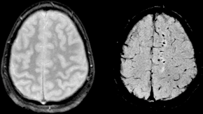
| Dua gambar MRI pasien dengan cedera aksonal difus akibat trauma, dengan kekuatan medan 1,5 tesla. Kiri: konvensional gradient recalled echo (GRE). Kanan: Susceptibility weighted image (SWI). | |
| Informasi umum | |
| Spesialisasi | Neurology |
Cedera aksonal difus (DAI) adalah cedera otak di mana lesi yang tersebar terjadi di area yang luas di saluran materi putih serta materi abu-abu.[1][2][3][4][5][6][7] DAI adalah salah satu jenis cedera otak traumatis yang paling umum dan merusak dan merupakan penyebab utama ketidaksadaran dan keadaan vegetatif persisten setelah trauma kepala parah. Ini terjadi pada sekitar setengah dari semua kasus trauma kepala parah dan mungkin merupakan kerusakan utama yang terjadi pada gegar otak. Hasilnya seringkali berupa koma, dengan lebih dari 90% pasien dengan DAI parah tidak pernah sadar kembali. Mereka yang terbangun dari koma seringkali tetap mengalami gangguan yang signifikan.
DAI dapat terjadi di seluruh spektrum keparahan cedera otak traumatis (TBI), di mana beban cedera meningkat dari ringan hingga berat.[8][9] Gegar otak mungkin merupakan jenis cedera aksonal difus yang lebih ringan.[9][10]
Referensi[sunting | sunting sumber]
- ^ Strich SJ (August 1956). "Diffuse degeneration of the cerebral white matter in severe dementia following head injury". Journal of Neurology, Neurosurgery, and Psychiatry. 19 (3): 163–85. doi:10.1136/jnnp.19.3.163. PMC 497203
 . PMID 13357957.
- ^ Povlishock JT, Becker DP, Cheng CL, Vaughan GW (May 1983). "Axonal change in minor head injury". Journal of Neuropathology and Experimental Neurology. 42 (3): 225–42. doi:10.1097/00005072-198305000-00002. PMID 6188807.
- ^ Adams JH (March 1982). "Diffuse axonal injury in non-missile head injury". Injury. 13 (5): 444–5. doi:10.1016/0020-1383(82)90105-X. PMID 7085064.
- ^ Christman CW, Grady MS, Walker SA, Holloway KL, Povlishock JT (April 1994). "Ultrastructural studies of diffuse axonal injury in humans". Journal of Neurotrauma. 11 (2): 173–86. doi:10.1089/neu.1994.11.173. PMID 7523685.
- ^ Povlishock JT, Christman CW (August 1995). "The pathobiology of traumatically induced axonal injury in animals and humans: a review of current thoughts". Journal of Neurotrauma. 12 (4): 555–64. doi:10.1089/neu.1995.12.555. PMID 8683606.
- ^ Vascak M, Jin X, Jacobs KM, Povlishock JT (May 2018). "Mild Traumatic Brain Injury Induces Structural and Functional Disconnection of Local Neocortical Inhibitory Networks via Parvalbumin Interneuron Diffuse Axonal Injury". Cerebral Cortex. 28 (5): 1625–1644. doi:10.1093/cercor/bhx058. PMC 5907353
 . PMID 28334184.
- ^ Smith DH, Hicks R, Povlishock JT (March 2013). "Therapy development for diffuse axonal injury". Journal of Neurotrauma. 30 (5): 307–23. doi:10.1089/neu.2012.2825. PMC 3627407
 . PMID 23252624.
- ^ Smith DH, Meaney DF (December 2000). "Axonal damage in traumatic brain injury". The Neuroscientist. 6 (6): 483–95. doi:10.1177/107385840000600611.
- ^ a b Blumbergs PC, Scott G, Manavis J, Wainwright H, Simpson DA, McLean AJ (August 1995). "Topography of axonal injury as defined by amyloid precursor protein and the sector scoring method in mild and severe closed head injury". Journal of Neurotrauma. 12 (4): 565–72. doi:10.1089/neu.1995.12.565. PMID 8683607.
- ^ Bazarian JJ, Blyth B, Cimpello L (February 2006). "Bench to bedside: evidence for brain injury after concussion--looking beyond the computed tomography scan". Academic Emergency Medicine. 13 (2): 199–214. doi:10.1197/j.aem.2005.07.031. PMID 16436787.
