
Berkas:SEM blood cells.jpg

Ukuran pratayang ini: 482 × 600 piksel. Resolusi lainnya: 193 × 240 piksel | 386 × 480 piksel | 617 × 768 piksel | 823 × 1.024 piksel | 1.800 × 2.239 piksel.
Ukuran asli (1.800 × 2.239 piksel, ukuran berkas: 1,33 MB, tipe MIME: image/jpeg)
Ringkasan
| Deskripsi |
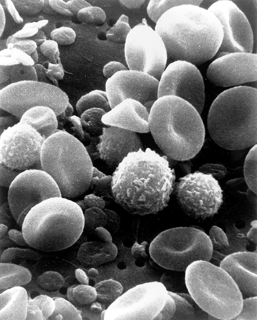
English: This is a scanning electron microscope image from normal circulating human blood. One can see red blood cells, several white blood cells including lymphocytes, a monocyte, a neutrophil, and many small disc-shaped platelets. Red cells are nonnucleated and contain hemoglobin, an important protein that contains iron and allows the cell to carry oxygen to other parts of the body. They also carry carbon dioxide away from peripheral tissue to the lungs where it can be exhaled. The infection-fighting white blood cells are classified in two main groups: granular and agranular. All blood cells are formed in the bone marrow. There are two types of agranulocytes: lymphocytes, which fight disease by producing antibodies and thus destroying foreign material, and monocytes. Platelets are tiny cells formed in bone marrow and are necessary for blood clotting. Type: Black & White Print Русский: Это изображение нормально циркулирующей крови человека получено с помощью сканирующего электронного микроскопа. Можно видеть красные кровяные тельца, несколько белых клеток крови (в их числе лимфоциты, моноциты, нейтрофилы) и множество мелких дискообразных пластинок. Красные кровяные тельца содержат гемоглобин — важный белок, который содержит железо и позволяет клетке переносить кислород к другим частям тела. Также они переносят углекислый газ от периферических тканей в лёгкие, где тот после газообмена может быть выдохнут. Лейкоциты борются с инфекциями и на две основные группы: гранулярные и агранулярные. Все клетки крови образуются в костном мозге. Есть два типа агранулоцитов: лимфоциты, которые борются с болезнью, производя антитела и тем самым разрушая чужеродный материал, и моноциты. Тромбоциты представляют собой крошечные клетки, образующиеся в костном мозге, и необходимы для свертывания крови. Тип фото: чёрно-белая печать. العربية : صورة بالمجهر الإلكتروني الماسح لدم الإنسان. يمكن للمرء أن يرى خلايا الدم الحمراء والعديد من خلايا الدم البيضاء بما في ذلك الخلايا الليمفاوية ووحيدات النوى والخلية المتعادلة والعديد من الصفائح الدموية الصغيرة ذات الشكل القرصي. |
||||||
| Tanggal | Date Created: February 1982 | ||||||
| Sumber | Image and description: National Cancer Institute | ||||||
| Pembuat | Bruce Wetzel (photographer). Harry Schaefer (photographer) | ||||||
| Izin (Menggunakan kembali berkas ini) |
|
||||||
| Versi lainnya |
Karya turunan dari berkas ini: |
||||||

| Annotations | This image is annotated: View the annotations at Commons |
Riwayat berkas
Klik pada tanggal/waktu untuk melihat berkas ini pada saat tersebut.
| Tanggal/Waktu | Miniatur | Dimensi | Pengguna | Komentar | |
|---|---|---|---|---|---|
| terkini | 3 Februari 2021 18.17 | | 1.800 × 2.239 (1,33 MB) | Tm | Reverted to version as of 20:27, 7 October 2006 (UTC) |
| 10 November 2020 04.50 |  | 1.800 × 2.239 (309 KB) | Ratmanz | Optimized. | |
| 7 Oktober 2006 20.27 |  | 1.800 × 2.239 (1,33 MB) | DO11.10 | ||
| 4 Oktober 2006 03.00 |  | 1.800 × 2.239 (989 KB) | DO11.10 | {{Information |Description=This is a scanning electron microscope image from normal circulating human blood. One can see red blood cells, several white blood cells including lymphocytes, a monocyte, a neutrophil, and many small disc-shaped platelets. Red | |
| 4 Oktober 2006 01.09 |  | 500 × 326 (36 KB) | DO11.10 | {{Information |Description= A three-dimensional ultrastructural image analysis of a T-lymphocyte (right), a platelet (center) and a red blood cell (left), using a Hitachi S-570 scanning electron microscope (SEM) equipped with a GW Backscatter Detector. |
Penggunaan berkas
5 halaman berikut menggunakan berkas ini:
Penggunaan berkas global
Wiki lain berikut menggunakan berkas ini:
- Penggunaan pada ar.wikipedia.org
- Penggunaan pada ar.wikiversity.org
- Penggunaan pada ast.wikipedia.org
- Penggunaan pada as.wikipedia.org
- Penggunaan pada az.wikipedia.org
- Penggunaan pada ba.wikipedia.org
- Penggunaan pada be-tarask.wikipedia.org
- Penggunaan pada be.wikipedia.org
- Penggunaan pada bg.wikipedia.org
- Penggunaan pada bn.wikipedia.org
- Penggunaan pada bn.wikibooks.org
- Penggunaan pada bs.wikipedia.org
- Penggunaan pada ca.wikipedia.org
- Penggunaan pada ce.wikipedia.org
- Penggunaan pada ckb.wikipedia.org
- Penggunaan pada cs.wikipedia.org
- Penggunaan pada cv.wikipedia.org
- Penggunaan pada cy.wikipedia.org
- Penggunaan pada de.wikipedia.org
- Penggunaan pada de.wikibooks.org
- Penggunaan pada dv.wikipedia.org
- Penggunaan pada el.wikipedia.org
- Penggunaan pada el.wiktionary.org
- Penggunaan pada en.wikipedia.org
Lihat lebih banyak penggunaan global dari berkas ini.